EKL coordinates project iAds – “Intelligent design of adenovirus vectors”.
iAds is a European Innovation Council (EIC) Pathfinder project that aims to maximise the potential of adenovirus vector for vaccines and gene transfer.
The consortium of 6 partners from 5 European countries will dedicate 4 years to develop an intelligent design of adenovirus vectors (iAds). The project aims to maximise the potential of adenovirus vectors for vaccines and gene transfer. The objective is to revolutionise gene transfer and generate solutions in areas of unmet medical need via a platform that exploits the full potential of viral vectors. iAds aims to target specific cell types in the heart and brain to improve gene therapy efficacy and safety.
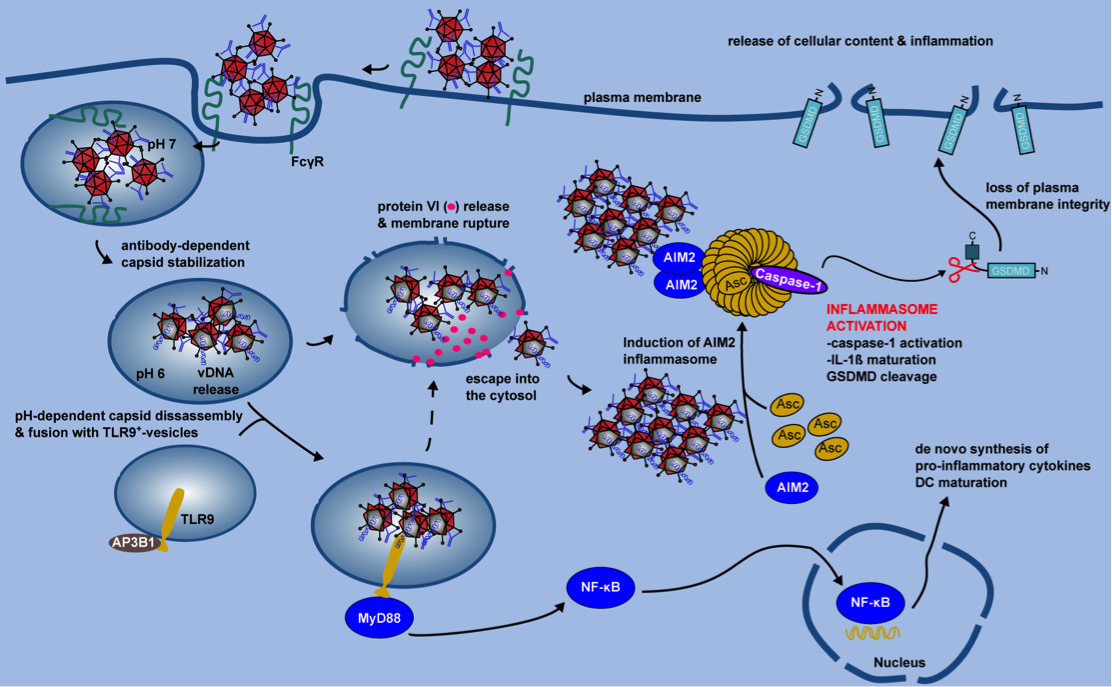
Gene therapy is a highly promising approach for treating certain diseases. Viral vectors have been developed stripped of their ability to cause disease and are instead used to deliver genetic material into cells for therapeutic purposes. Adenoviruses are particularly attractive because they can efficiently deliver DNA into both dividing and non-dividing cells. Funded by the European Innovation Council, the iAds project aims to overcome the limitations of adenoviral vectors, such as host immune responses and imperfect targeting. The consortium will create an in-silico platform for the design of intelligent adenovirus vectors with a focus on heart- and brain-specific targeting, addressing areas of unmet medical need.
For more information: www.intelligentadenoviruses.eu
DSynchro ANR-21-CE17-0056 CE17 Translational Research
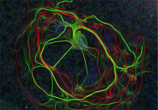
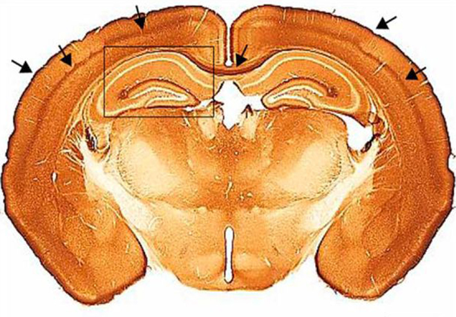
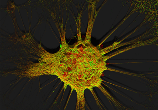
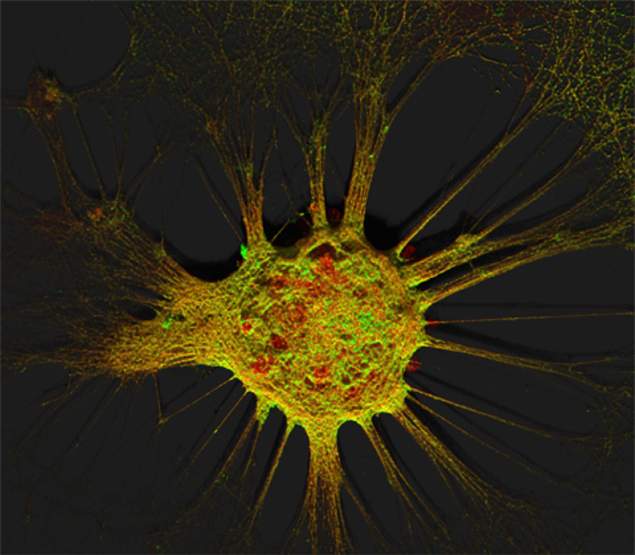
Targeting exogenous NaV1.1 activity to inhibitory neurons in mice with Dravet syndrome
There are 80 billion neurons in the human brain. To determine which population and location in the brain requires modification to help children with Dravet syndrome, further research is necessary. Dravet syndrome is a severe epilepsy that affects infants. It is caused by a genetic mutation in the SCN1A gene, which encodes a sodium channel (NaV1.1) responsible for transporting positively charged sodium ions into cells, particularly neurons. When NaV1.1 function is impaired, it disrupts the balance between excitation and inhibition in the brain, leading to various complications including seizures. While conventional medications can reduce seizures, they have limited impact on developmental delays. Therefore, novel therapeutic approaches are required to assist children with Dravet syndrome.
Gene therapy, which employs DNA as a therapeutic agent to address the underlying cause of the disease rather than its symptoms, holds potential for treating Dravet syndrome. To achieve this, precise vector design and targeted delivery to the affected neurons are essential. The primary challenges in treating neurodevelopmental diseases with gene therapy include:
- Identifying the appropriate cell types (neurons, astrocytes and microglia) in the correct brain regions.
- Correcting the cellular functions.
- Sustaining the survival of healthy cells in areas potentially damaged by the disease.
- Modifying a sufficient number of cells to produce a meaningful therapeutic effect.
- Ensuring the long-term efficacy of the treatment.
For the past decade, our research has focused on developing a therapy for Dravet syndrome. However, we have not prioritised the inhibitory neuron population, which is considered the most critical for the condition. Despite this, we observed improvements in reducing seizures and extending the lifespan of mice with Dravet syndrome.
DSynchro is focused on advancing therapy by identifying which neurons require increased NaV1.1 expression. Our consortium was uniquely positioned to develop a novel treatment. We discovered that, contrary to the prevailing theory, NaV1.1 is produced by most neuron types and that correcting the SCN1A gene in multiple neuron types is necessary to treat seizures and restore the balance between excitation and inhibition.
We are sharing our findings with scientists, clinicians and entrepreneurs through publications* and presentations. This has resulted in the establishment of a CNRS-led spin-off in 2024 to treat Dravet syndrome.
*Fadila S, Beucher B, Dopes-Reyes IG, Mavashov A, Brusel M, Anderson K, Ismeurt C, Goldberg EM, Ricobaraza A, Hernandez-Alcoceba R, Kremer EJ & Rubinstein M. Viral vector-mediated delivery of SCN1A reduces Dravet syndrome seizures in juvenile and adolescent mice. J Clin Invest. 2023;133(12):e159316; doi:10.1172/JCI159316.
*Fadila S, Krivoshein G, Majadly H, Mavashov A, Ranen S, Brusel M, González Dopeso-Reyes I, Beucher B, Kremer EJ, Tolner E M Rubinstein. Disrupted temperature-sleep coupling mechanism in a Dravet syndrome mouse model. Nature Comm 17, 3232 (2026) doi: 10.1038/s41467-026-69957-1